
Histology and Cellular Imaging
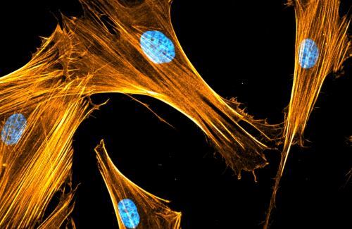
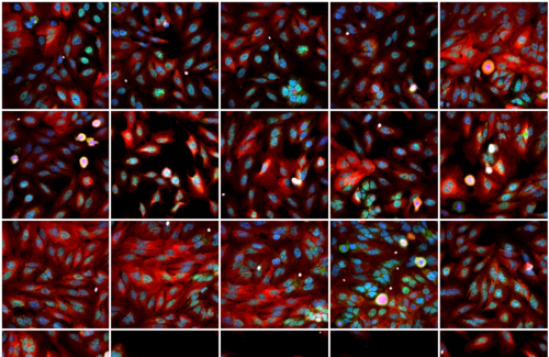
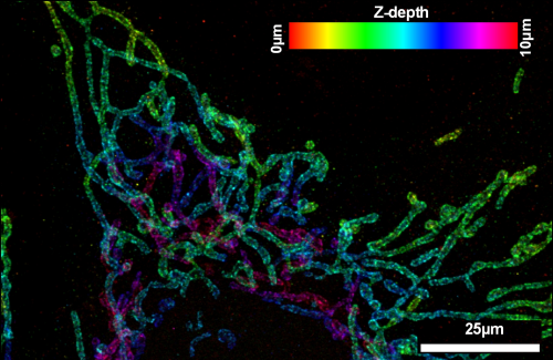
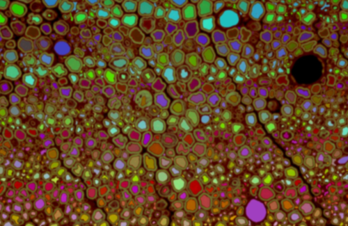
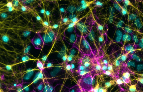
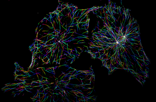
The Histology and Cellular Imaging (HiCI) unit provides microscopy-related support for scientific projects in the VIB-UAntwerp Center for Molecular Neurology. Experience and infrastructure is present for sample preparation (histology, immunocyto- and histochemistry, live cell imaging, electron microscopy, superresolution microscopy, ..) and for basic and advanced microscopic imaging, either via in-house equipment or via a network of external (VIB or UAntwerp) collaborations.
Employed imaging modalities include widefield fluorescence; laser scanning and spinning disk confocal microscopy; scanning, transmission and volume electron microscopy; expansion microscopy; automated high content/throughput microscopy, … Image and data analysis is implemented through customized project-driven bio-image informatical pipelines. Example projects can be found here. If you have questions, or requests, please contact dr. Bob Asselbergh
Our expert
Bob Asselbergh
Research Associate, Microscopy Expert